

STREXS是一种模拟细胞生长过程中机械应力的装置。张力腔室中的细胞被仪器拉伸或压缩。施加在细胞上的机械应力更好的模拟了自然动态生理环境,可精准控制施加在细胞上的应力级别。
细胞应力拉伸仪(STREX)的优势
× 精准的应力程序设计
× 高频率和低频率处可实现连续拉伸/压缩
× 培养腔室所受应力的均一性
× 横向剪切力非常小
× 操作便捷
| 型号 | ||||
| 项目 | ST-140-04 | ST-140-10 | ST-150 | ST-190-XY |
| 应用范围 | ? 细胞骨架重组 ? 细胞形态学 ? 基因或蛋白表达 ? 信号传导 ? 长期性研究(几小时到几天) | ? 细胞骨架重组 ? 细胞形态学 ? 离子调控 ? 钙离子流入 ? 氮氧化合物的合成 ? 培养过程的实时监测 ? 短时间的过程研究(15-20min) | ||
| 腔室数目 | 6 | 5 | 1 | 1 |
| 腔室大小—培养面积 | 4cm2 | 10cm2 | 4cm2 | 4cm2 |
| 应力方向 | 单轴拉伸 | 单轴拉伸 | 单轴拉伸 | 双轴拉伸/压缩 |
| 与显微镜兼容性 | - | - | 兼容Nikon, Olympus, 可选Zeiss, Leica | 兼容Nikon, Olympus, 可选Zeiss, Leica |
| 培养箱 | 标准培养箱 | 标准培养箱 | 标准培养箱 | 标准培养箱 |
| 程序个数 | 64 | 64 | 64 | 64 |
The exceptional physical and chemical properties of the silicone elastomer PDMS (polydimethylsiloxane) create a specially flexible thin-membrane chamber.
● High Reproducability:
Springy PDMS chambers bounce back from stretching and compression with their original properties intact. Thus, the chambers demonstrate good reproducibility in applications requiring continuous mechanical stretching over prolonged periods.
● Superior Transparency:
An optically transparent, ultra-thin (100-200 µm) membrane at the well bottom not only makes stretch chambers compatible with optical microscopy techniques, but with fluorescence detection and microscopy as well.
Uniformity of direction and force are issues of crucial concern in cell stretching. Stretching systems and chambers with insufficient properties can cause extraneous stretching on the wrong axis and generate a secondary load in that direction. In addition, these systems may not apply stress equally to all cells in the chamber, making it impossible to accurately gauge the effect of the stretch stimulus across the unevenly treated sample.
The STREX Cell Stretching System is designed to achieve stretching in a single, parallel direction, with only a very weak secondary load. Research has demonstrated that the STREX system enables highly reproducible cyclic stretching over prolonged periods at ratios of 1-20%.(Ref)
Because the methyl groups align themselves to the surface, the stretch chamber is highly water-repellent, meaning that cells will not adhere without pre-treatment of the chamber. Thus, the PDMS chamber surface needs to be coated with extracellular matrix in order to successfully adhere and culture cells. Fibronectin, collagen, gelatin and laminin, among others, can be used for this purpose.
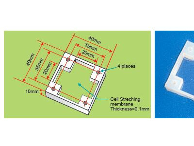
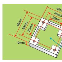
STREX offers standard stretch chambers in two sizes, the 4cm² and 10cm² models. Specialized versions are also available, including multi-well, gel-stretching, and transwell experiment chambers.
(Ref) Naruse, K., Yamada, T., Sai, X.R., Hamaguchi, M., Sokabe, M. Pp125FAK is required for stretch dependent morphological response of endothelial cells. Oncogene 1998, 17:455-463.
Stretch Chambers
STB-CH-04 Effective size:2.0×2.0×1.0cm
| Compatible with STB-10-04 STB-140-04 STB-150 STB-195 |
| Catalog No. STB-CH-04 | Item name 4cm² chamber (10 pcs.)/1 set |
STB-CH-10 Effective size: 3.2×3.2×1.0cm
| Compatible with STB-10-10 STB-140-10 |
| Catalog No. STB-CH-10 | Item name 10cm² chamber (5 pcs.)/1 set |
STB-CH-04-XY
Compatible with
XY-biaxial stretching Effective size: 2.0×2.0×1.0cm
| Compatible with STB-190-XY |
| Catalog No. STB-CH-04-XY | Item name 4cm² XY chamber (5 pcs.)/1 set |
STB-CH-4W multiwell type Effective size: 1.5×1.5×1.0cm
| Compatible with STB-10-10 STB-140-10 |
| Catalog No. STB-CH-4W | Item name 10cm² chamber with 4-wells (5 pcs.)/1 set |
Note: Chamber specifications:
Heatproof temperature: 5℃-180℃, Humidity: 20-100%
Durability: 900,000cycles approx. (when stretching ratio=20%)
Note: Store unused chambers in a cool dark place. Chambers are denatured by UV-rays irradiation.
Chamber Stands
STB-CH-04ST-XX chamber stands
Note: "XX" denotes stretching ratio. ex)STB-CH-10ST-05: for use with 5% stretching ratio
Stretching stands for STB-CH-04.0
| Compatible with holds 1 chamber, STB-CH-04 |
| Catalog No. STB-CH-04ST-XX | Item name stands for 4cm² chambers | Price Please contact us |
STB-CH-10ST-XX chamber stands
Note: "XX" denotes stretching ratio. ex)STB-CH-10ST-05: for use with 5% stretching ratio
Stretching stands for STB-CH-10.0
| Compatible with holds 1 chamber, STB-CH-10 |
| Catalog No. STB-CH-10ST-XX | Item name Stands for 10cm² chambers | Price Please contact us |
Coating Protocols for Stretch Chambers
Stretch chambers as sold are not sterile and provide no cell adhesion. Therefore, the chamber must first be autoclaved for 20 minutes at 121° C, and then the membrane must be coated with extracellular matrix before seeding cells.
Coating protocol: Fibronectin
- Prepare a fibronectin solution by dissolving 0.05 mg/ml of fibronectin in PBS.
- Place the stretch chamber in a culture dish and pour the fibronectin solution into the chamber well so that it completely covers the bottom surface.
- Incubate the fibronectin-treated chamber in the culture dish at 37° C for at least four hours.
- Remove the culture dish from incubator, and draw up any remaining solution from the chamber using a pipette or other suitable device.
Coating Protocol: Collagen
- Prepare and autoclave a dilution of hydrochloric acid (pH3.0, 1mM).
- Dilute type 1 collagen in the autoclaved hydrochloric acid.
- Place the stretch chamber in a culture dish and pour the collagen solution into the chamber well so that it completely covers the bottom surface.
- Cover the culture dish with a lid and incubate at 37° C for at least four hours.
- Remove the culture dish from incubator, and leave to stand for a period. Then draw up the remaining solution from the chamber using a pipette or other suitable device.
- Rinse the chamber twice with serum-free culture fluid to remove any excess collagen solution that may have remained after the above steps.
Coating protocol: Gelatin
- Prepare a gelatin solution by dissolving 2% gelatin powder in PBS. Autoclave the gelatin solution
- Place the stretch chamber in a culture dish and pour the gelatin solution into the chamber well so that it completely covers the bottom surface.
- Incubate the gelatin-treated chamber in the in the culture dish at 37° C for at least four hours.
- Remove the culture dish from incubator, and draw up any remaining solution from the chamber using a pipette or other suitable device.
Protocol of Fluorescent Staining after Stretching
- Splice out cell plane from stretch chamber with surgical knife.
(as large as approx. 0.8×0.5cm)->the size varies according to the size of container to be used since step 2.
Splicing to make the longer side get along with the stretching direction makes it easy to find out the stretching direction afterwards.
- put the membrane spliced in step 1 into the container with PBS(‐).
(using 1×1 chamber(without frame) attached on a cover glass.) - PBS(‐) wash, 2 times
- fix with 4% formalin solution in PBS(‐)->RT, 5 min, shake
- PBS(‐) wash, RT, 5 min, 3 times
- 0.1% TritonX-100 in PBS(‐), pierce cell membrane->RT, 5 min, shake
- PBS(-) wash->RT, 5 min, 3 times
- block 2% BSA in PBS(‐)->RT, 30 min, shake
- primary antibody in 0.1%Tween20 in PBS(‐)->RT, 30min, shake
- 0.1%Tween20 in PBS(‐) wash->RT, 5 min, 3 times
- secondary antibody in 0.1%Tween20 in PBS(‐)->RT, 30 min, shake
- 0.1%Tween20 in PBS(‐) wash->RT, 5 min, 3 times
- put on a slide glass to enclose(Perma Fluor)->put the cell membrane side down
- Fluorescent observation