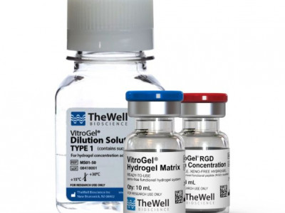
在The Well Bioscience,我们相信每个人都是独特的,个性化的药物可以改善每个患者的生活质量。我们专注于使用3D生物塑料平台来推动先进的生物医学,
用于精密医学,细胞疗法和生物处理。作为无XENO 3D细胞平台的先驱,我们模仿人类的微观环境,以进行器官形成,干细胞缩放和智能细胞输送系统。
通过与学术界,医院和制药行业的科学家合作,我们旨在改善全球患者的生物医学。
我们的无XENO水凝胶系统(Vitrogel®)可以紧密模仿天然的细胞外基质,并为3D类器官模型的临床应用打开门。来自不同来源(例如患者衍生细胞)
的细胞可以使用我们的系统进行培养,该系统具有完全实验室的自动化潜力的个性化3D细胞模型,包括高度复杂的类器官模型和共培养系统。
我们的技术可以通过将其用于组织工程医疗产品的3D生物打印,可注射的辅助物,以实现更好的细胞保留率和更高的细胞活力来进行细胞治疗,
以及用于大规模细胞/生物生物生物生产制造的3D干细胞量表系统。
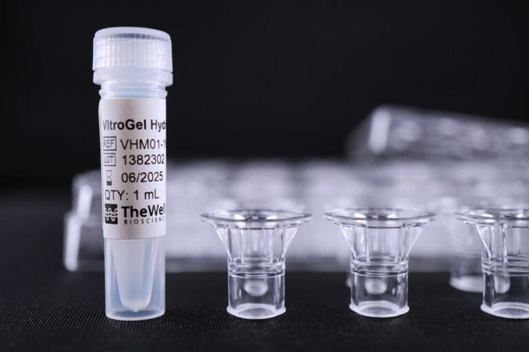
VitroGel细胞侵袭试剂盒,无异源水凝胶系统,用于细胞侵袭的一致研究
【产品介绍】
VitroGel-Based Cell Invasion Assay Kits 无异源水凝胶系统,用于细胞侵袭研究。
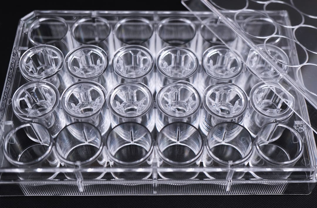
VitroGel细胞侵袭试剂盒由 VitroGel® 水凝胶(多功能、无异种物质、生物功能水凝胶,紧密模仿生理细胞外基质)与优质 VitroPrime™ 细胞培养插入物相结合,可实现更准确和一致的侵袭和迁移研究,优于动物来源 ECM。
即用型 VitroGel细胞侵袭试剂盒可以进行传统细胞侵袭检测研究。
任何可调节的 VitroGel 高浓度细胞侵袭试剂盒,其生物物理和生化特性均可调节,使研究人员能够探索不同的基质强度、配体、趋化因子、生长因子等如何影响细胞侵袭。
无论是使用即用型 VitroGel 水凝胶还是任何可调节的高浓度 VitroGel 水凝胶,都可以用于细胞侵袭实验,为细胞迁移研究提供多功能性。
【产品规格】
Use使用 | 细胞侵袭、细胞迁移、共培养、屏障模型 |
Inserts | 8 µm,PET,透明,无菌。 12 个插入物/板。 |
即用型试剂盒 | VitroGel 水凝胶+ VitroPrime 细胞培养插入物 IA-VHM01-1P IA-VHM01-4P |
高浓度试剂盒 | VitroGel 高浓度水凝胶 + VitroPrime 细胞培养插入物 IA-HC001-1P, IA-HC001-4P IA-HC003-1P, IA-HC003-4P IA-HC007-1P, IA-HC007-4P IA-HC008-1P, IA-HC008-4P IA-HC009-1P, IA-HC009-4P IA-HC010-1P, IA-HC010-4P |
成分 | 无异源的功能性水凝胶 |
pH | 中性 |
使用次数 | 1 mL 水凝胶/12 个插入物 |
贮存 | 2-8°C 储存。室温运输。 |
【产品特点】
• 调节 ECM 硬度和成分 – 控制关键化合物并探索不同的基质强度、配体、趋化因子、生长因子等如何影响细胞侵袭。
• 批次间的一致性——体系成分明确,与动物源性 ECM 不同,没有未知蛋白质成分。
• 无需冰上操作——室温稳定,可在 30 分钟内完成,并可适应实验室自动化。
• 支持屏障模型——该体系可用于创建体外复杂的微环境,例如人体肠道模型。
【VitroGel即用型细胞侵袭试剂盒、高浓度细胞侵袭试剂盒与其他动物来源ECM比较】
【Vitrogel 细胞侵袭试剂盒操作流程】
1、使用基于 VitroGel 的细胞侵袭试剂盒,不仅可以进行传统的侵袭/迁移检测,还可以使用可调节试剂盒研究更多类型的侵袭/迁移研究。

2、细胞侵袭培养过程仅需30分钟 “Just add cells”
无需冰上操作,在室温下完成实验。基于 VitroGel 的细胞侵袭检测试剂盒即用型。不需要交联剂。

产品介绍】
VitroGle® Organoid Recovery Solution是一款无酶的细胞回收液,可15 min内快速的从VitroGel或动物源ECM系统上高效率地回收类器官/细胞。
【产品规格】
货号 | MS04-100, MS04-500 |
|---|---|
| 规格 | 100 mL, 500 mL |
| 配方 | 不含酶 |
| 用途 | 收获从动物源ECM或 VitroGel水凝胶培养的类器官/细胞 用于样本固定、染色成像或下游数据分析 |
| 时间 | 10-15 min |
| 下游应用 | 回收的细胞可以再就行3D和2D培养 |
| pH | 中性 |
| 储存 | 2-30 °C |
| 稳定性 | 12 个月 |
| 运输 | 室温 |
【产品特点】
▪ 两分钟从动物源ECM上回收完整的类器官/细胞
▪ 高回收率
▪ 支持从2D ECM包被平板上回收细胞
▪ 稳定的无酶配方
▪ 易于使用,室温下操作
▪ 保质期长,无需冷藏包装运输
【VitroGel类器官回收液与其它品牌回收液的比较】
| 项目 | VitroGel | Company C | Company R | Company S |
| 从动物源ECM回收的时间 | 2 min | 约60 min | 约60 min | 至少30 min |
| 回收率&细胞活率 | 高 | 中 | 中 | 中 |
| 便捷性 | 高 | 低 | 低 | 中 |
| 从2D ECM包被平板回收细胞 | 可以 | / | / | / |
| 无需冷链运输 | 是的 | / | / | 是的 |
| 存储条件 | 2-30℃ | 2-8℃ | 2-8℃ | 15-35℃ |
| 保质期 | 12个月 | 3个月 | 2个月 | N/A |
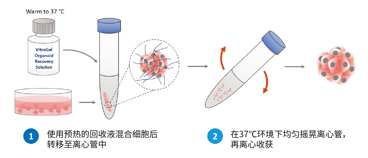
【类器官回收液操作流程】
1、动物源ECM系统的类器官/细胞回收操作
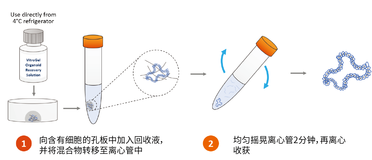
2、VitroGel系统的类器官/细胞回收操作
【类器官回收液应用比较】
1、VitroGel类器官回收液回收Matrigel系统上类器官两种方法的效果比较
A、Method 1:利用VitroGel类器官回收液重悬类器官培养物,移液吹打形成小块进行传代培养/扩增。
B、Method 2:在不使用移液管的前提下,利用VitroGel类器官回收液混合类器官培养物,均匀摇晃离心管以收获完整的类器官。
结果显示,两种方法均可以回收获得较为完整的类器官,且传代培养后正常增殖
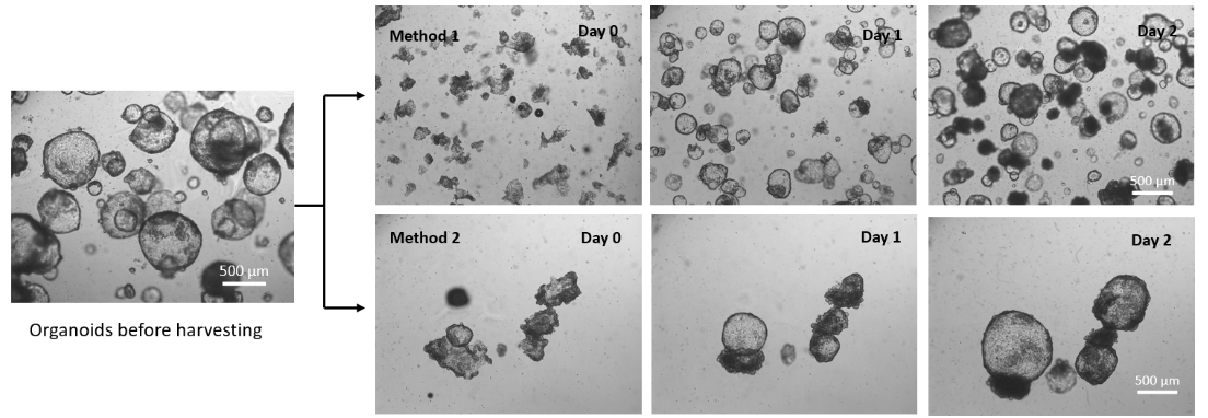
图1 VitroGel类器官回收液回收Matrigel系统上类器官两种方法的效果比较
2、VitroGel类器官和细胞回收液回收Matrigel系统上类器官的效果比较 在Matrigel上培养类器官3天,通过使用VitroGel类器官回收液和VitroGel细胞回收液进行Matrigel类器官的回收。
结果显示,室温下分别孵育2分钟(VitroGel类器官回收液,MS04)和10分钟(VitroGel细胞回收液,MS03),离心收集类器官后,MS04溶液中的Matrigel完全溶解,
而MS03溶液中有少量的软凝胶残留。两种回收液收获的类器官继续在Matrigel上传代培养,下图显示了良好的类器官扩增效果。
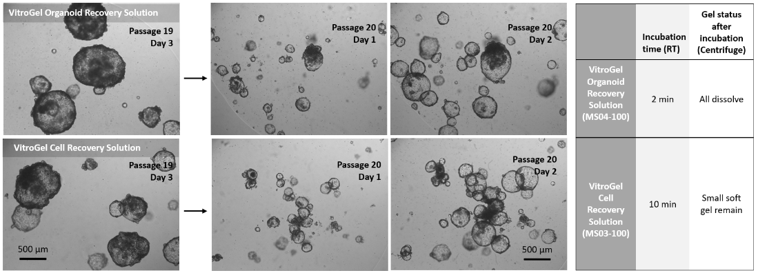
图2 VitroGel类器官和细胞回收液回收Matrigel系统上类器官的效果比较
3、VitroGel类器官回收液与其它品牌类器官回收液的效果比较 利用MS04溶液与其它品牌的类器官回收液(SC公司的RLR溶液、SA公司的3OHS)开展对比研究。
结果显示,MS04溶液可轻松回收类器官,并破碎成小碎片状进行传代培养;而RLR溶液和3OHS溶液回收的类器官很难破碎成小块状,且凝胶残留较严重。MS04溶液显示了更高的类器官回收率。

图3 VitroGel类器官回收液与其它品牌类器官回收液的效果比较
| 项目 | VitroGel类器官回收液 | SC公司RLR溶液 | SA公司3OHS溶液 |
| 孵育时间 | 2 min(室温) | >15 min(冰上) | >15 min(冰上) |
| 胶的溶解效果(离心) | 全部溶解 | 大部分凝胶残留 | 大部分凝胶残留 |
| 摇晃重悬为小块类器官 | 容易 | 困难 | 困难 |
| 回收效率 | 高 | 低 | 低 |
4、VitroGel类器官回收液回收Matrigel 2D包被培养的iPSC
使用MS04溶液回收Matrigel上培养的iPSC细胞,细胞脱落效果良好(图A),培养孔板上基本没有细胞残留(图B),再次接种后细胞生长状态良好(图C)。

图4 VitroGel类器官回收液回收Matrigel 2D包被培养的iPSC
(A、细胞脱落的形态;B、细胞脱落后的平板;C、传代后细胞形态)
【小结】
目前,类器官培养技术正处于技术爆发和科研成果井喷的阶段,行业发展具有很大的前景,但挑战也是并存的。例如,大多数类器官本身不具备血管化的结构;如何在体外模拟肿瘤类器官与免疫细胞相互作用关系;类器官的重复性和一致性;以及类器官下游应用的准确性等。总之,新的技术不断涌现,必然助力再生医学与精准医疗领域的研究更上一层楼。
| 产品名称 | 产品货号 | 规格 | 产品描述 |
|---|---|---|---|
| VitroGel® Organoid | VHM04-K | 4*2 mL | 即用型,类器官培养水凝胶,适用于多种类器官培养 |
| VitroGel® Cell Recovery Solution | MS03 | 100 mL | 无酶配方细胞/类器官回收液,从多种凝胶系统回收细胞/类器官 |
| VitroGel® Organoid Recovery Solution | MS04 | 100 mL |
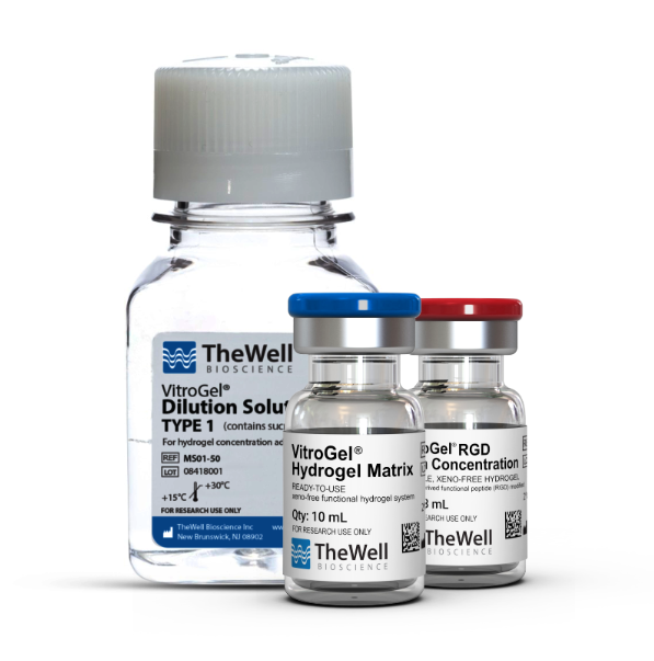
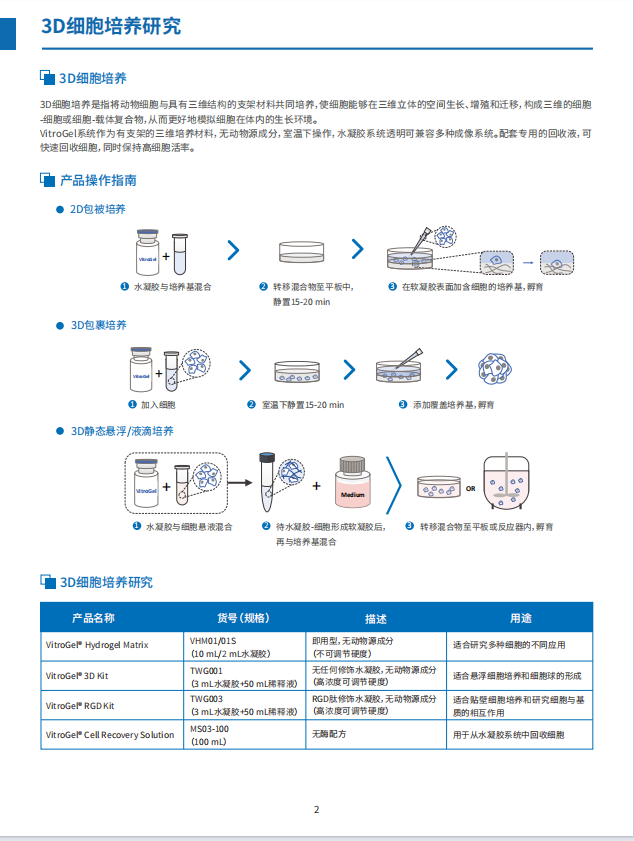
VitroGel™3D为无动物源性多糖水凝胶体系,模仿天然细胞外基质(ECM)环境, 只需室温操作即可。
工作原理
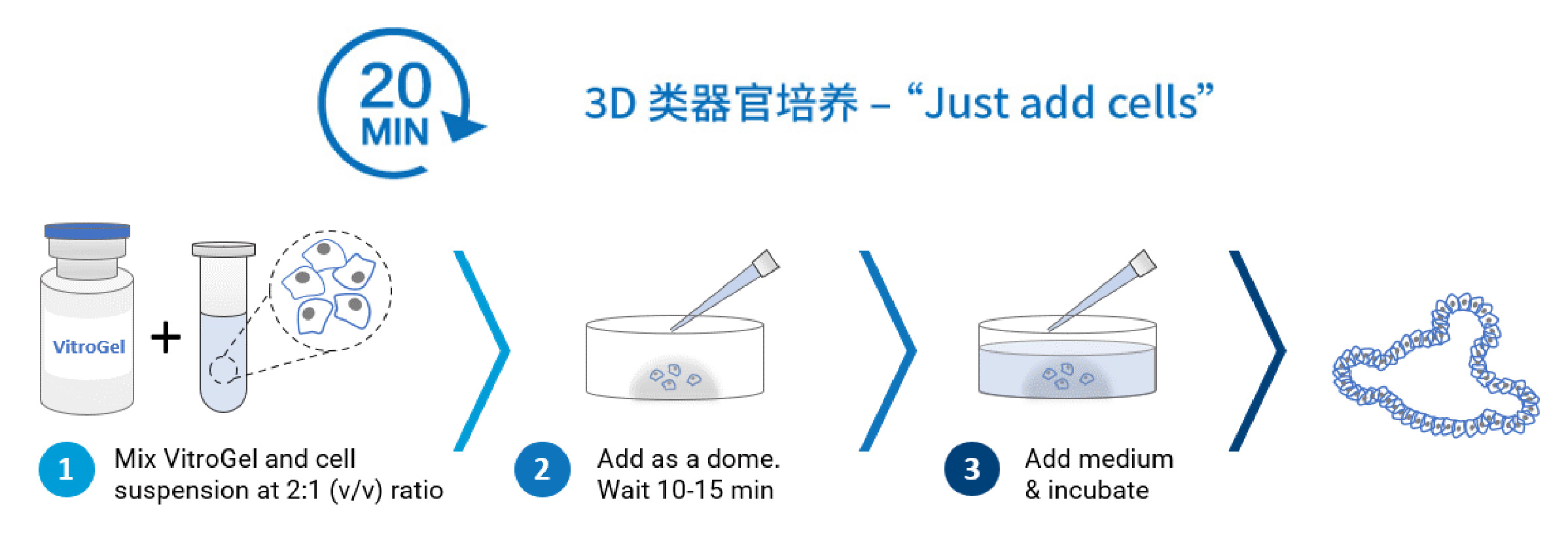
20分钟快速成胶(包括10-15分钟水凝胶稳定时间)

产品特点
即用型
无需特殊交联试剂,只需调整后与细胞混匀,添加培养基并培养即可。
可调节系统
可以通过调节不同水凝胶强度,为细胞创造更优生长微环境。
Xeno-free
多聚糖水凝胶,无任何动物源成分
室温操作
水凝胶可在室温条件下稳定保存操作,无需冰上操作
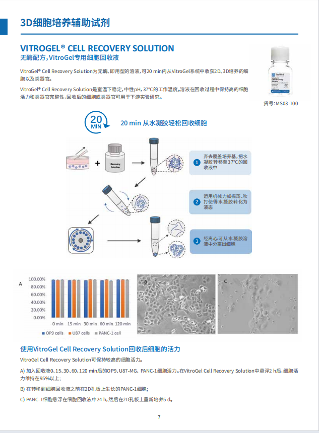
快速细胞收获- 20min操作方案
3D细胞培养后,可使用我们的无酶细胞回收液,从水凝胶中安全快速收获细胞,同时保持高细胞活力。
混合搭配
通过将不同类型的VitroGel混合使用,可构建多功能水凝胶。
透明
水凝胶颜色透明的,与不同的成像系统相容,便于细胞观察。
可注射
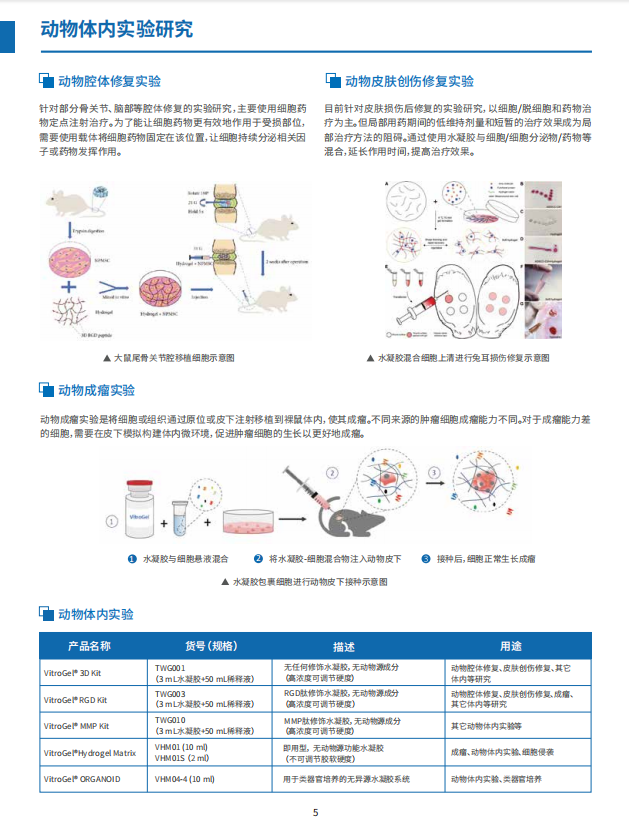
在软水凝胶形成后,可用于动物体内注射,研究细胞治疗和药物传递!
产品应用
3D细胞培养(3D Cell Model)
共培养(Co-Culture)
类器官培养(Organoid)
细胞侵袭实验(Invasion Assay)
神经细胞培养(Neuron)
干细胞培养
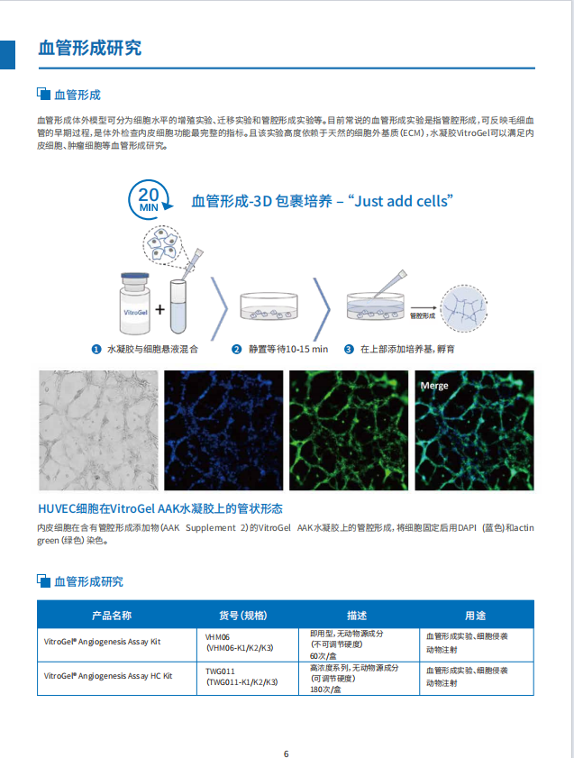
血管生成试验(Angiogenesis Assay)
动物体内注射(In vivo Injection)
订购信息
VitroGel系统可以相互混合搭配使用,可以通过混合不同的VitroGel系统来量身定制3D培养微环境。
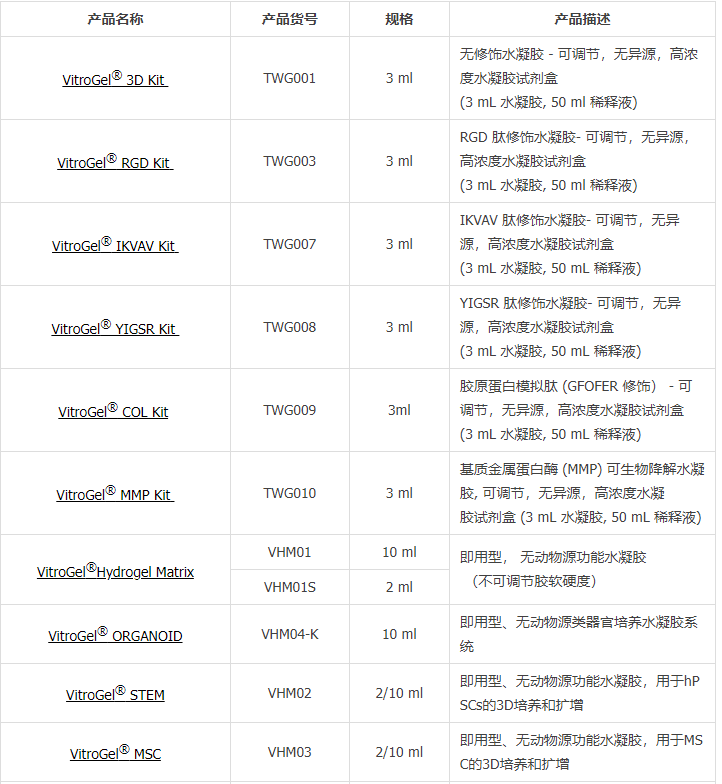
| 产品名称 | 产品货号 | 规格 | 产品描述 |
| TWG001 | 3 ml | 无修饰水凝胶 - 可调节,无异源,高浓度水凝胶试剂盒 (3 mL 水凝胶, 50 ml 稀释液) | |
| TWG003 | 3 ml | RGD 肽修饰水凝胶- 可调节,无异源,高浓度水凝胶试剂盒 (3 mL 水凝胶, 50 ml 稀释液) | |
| TWG007 | 3 ml | IKVAV 肽修饰水凝胶- 可调节,无异源,高浓度水凝胶试剂盒 (3 mL 水凝胶, 50 mL 稀释液) | |
| TWG008 | 3 ml | YIGSR 肽修饰水凝胶- 可调节,无异源,高浓度水凝胶试剂盒 (3 mL 水凝胶, 50 mL 稀释液) | |
| TWG009 | 3ml | 胶原蛋白模拟肽 (GFOFER 修饰) - 可调节,无异源,高浓度水凝胶试剂盒 (3 mL 水凝胶, 50 mL 稀释液) | |
| TWG010 | 3 ml | 基质金属蛋白酶 (MMP) 可生物降解水凝胶, 可调节,无异源,高浓度水凝 胶试剂盒 (3 mL 水凝胶, 50 mL 稀释液) | |
| VHM01 | 10 ml | 即用型, 无动物源功能水凝胶 (不可调节胶软硬度) | |
| VHM01S | 2 ml | ||
| VHM04-K | 10 ml | 即用型、无动物源类器官培养水凝胶系统 | |
| VHM02 | 2/10 ml | 即用型、无动物源功能水凝胶,用于hPSCs的3D培养和扩增 | |
| VHM03 | 2/10 ml | 即用型、无动物源功能水凝胶,用于MSC的3D培养和扩增 | |
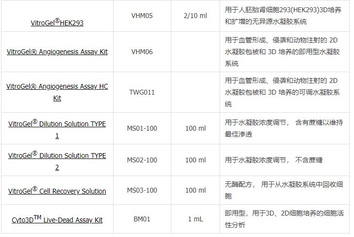
| VHM05 | 2/10 ml | 用于人胚胎肾细胞293(HEK293)3D培养和扩增的无异源水凝胶系统 | |
| VHM06 | 用于血管形成、侵袭和动物注射的 2D 水凝胶包被和 3D 培养的即用型水凝胶系统 | ||
| TWG011 | 用于血管形成、侵袭和动物注射的 2D 水凝胶包被和 3D 培养的可调水凝胶系统 | ||
| MS01-100 | 100 ml | 用于水凝胶浓度调节, 含有蔗糖以维持最佳渗透 | |
| MS02-100 | 100 ml | 用于水凝胶浓度调节, 不含蔗糖 | |
| MS03-100 | 100 ml | 无酶配方, 用于从水凝胶系统中回收细胞 | |
| BM01 | 1 mL | 即用型,用于3D、2D细胞培养的细胞活性分析 |
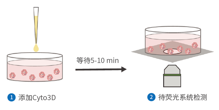
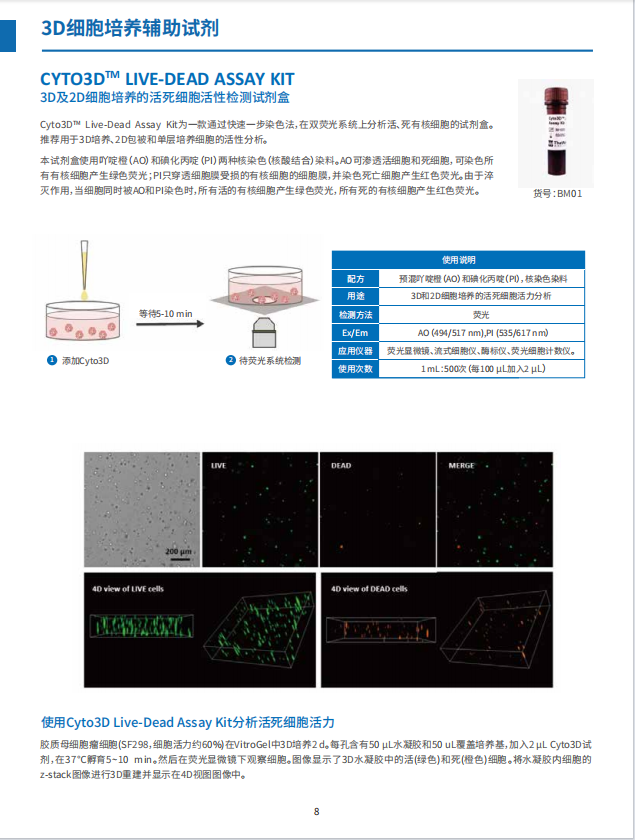
Cyto3D™ Live-Dead Assay Kit为一款通过快速一步染色,在双荧光系统上分析活、死有核细胞的试剂盒。推荐用于3D培养、2D包被和单层培养细胞的活性分析。
货号
BM-01S
工作原理
即用型
快速
灵敏
适用于3D细胞培养
成本低
本试剂盒使用吖啶橙(AO)和碘化丙啶(PI)两种核染色(核酸结合)染料。AO可渗透活细胞和死细胞,可染色所有有核细胞产生绿色荧光;PI只穿透细胞膜受损的有核细胞的细胞膜,并染色死亡细胞产生红色荧光。由于猝灭作用,当细胞同时被AO和PI染色时,所有活的有核细胞产生绿色荧光,所有死的有核细胞产生红色荧光。
使用说明 | |
配方 | 预混吖院橙(AO)和碘化丙(PI)核染色染料 |
|---|---|
用途 | 3D和2D细胞培养的活死细胞活力分析 |
检测方法 | 荧光 |
Ex/Em | AO(494/517 nm)PI(535/617 nm) |
应用仪器 | 荧光显微镜、流式细胞仪、酶标仪、荧光细胞计数仪 |
使用次数 | 1mL:500次(每100 μL加入2 μL) |
工作方法

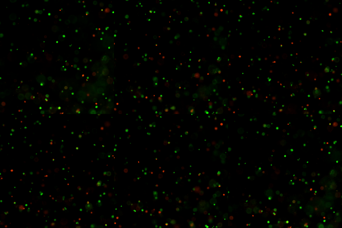
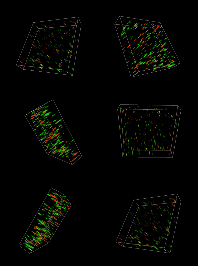
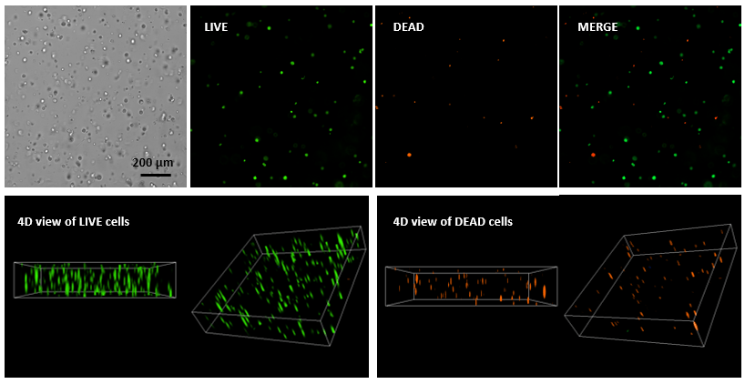
igure 1. Live-dead cell viability analysis by using Cyto3D Live-Dead Assay Kit.
Glioblastoma cells (SF 298, about 60% cell viability) were 3D cultured in VitroGel system for 2 days. 2 µL of Cyto3D reagent was added to each well containing 50 µL hydrogel and 50 µL cover medium. The mixture was incubated at 37 °C for 5-10 min. The cells were then observed under a fluorescent microscope. The images show the Live (green) and Dead (orange) cells within the 3D hydrogel matrix. The z-stack images of cells within hydrogel were then 3D reconstructed and showed in the 4D view images. The live and dead cells at higher levels of the hydrogel clearly show in the images by using Cyto3D Live-Dead Assay Kit.
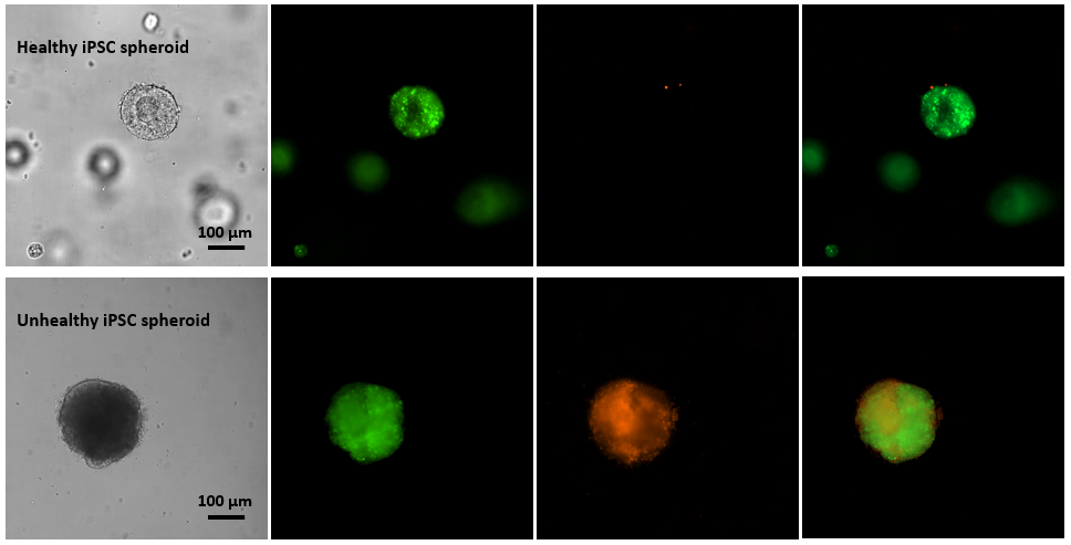
Figure 2. Live-dead cell viability images of stem cell spheroids.
Stem cells were static suspension-cultured in VitroGel STEM (CAT# VHM02) for 5 days. 2 µL of Cyto3D reagent was added to each well containing 100 µL cell suspension. The mixture was incubated at 37 °C for 5-10 min. The cells were then observed under a fluorescent microscope. The images show the Live (green) and Dead (orange) stem cell spheroids cultured in a 3D hydrogel matrix. The live-dead dyes of Cyto3D Live-Dead Assay Kit can successfully penetrate into large cell spheroids for cell viability analysis.
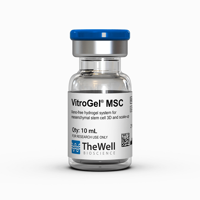
ready-to-use, xeno-free (animal origin-free) hydrogel system for mesenchymal stem cell 3D culture and scale-up
Overview
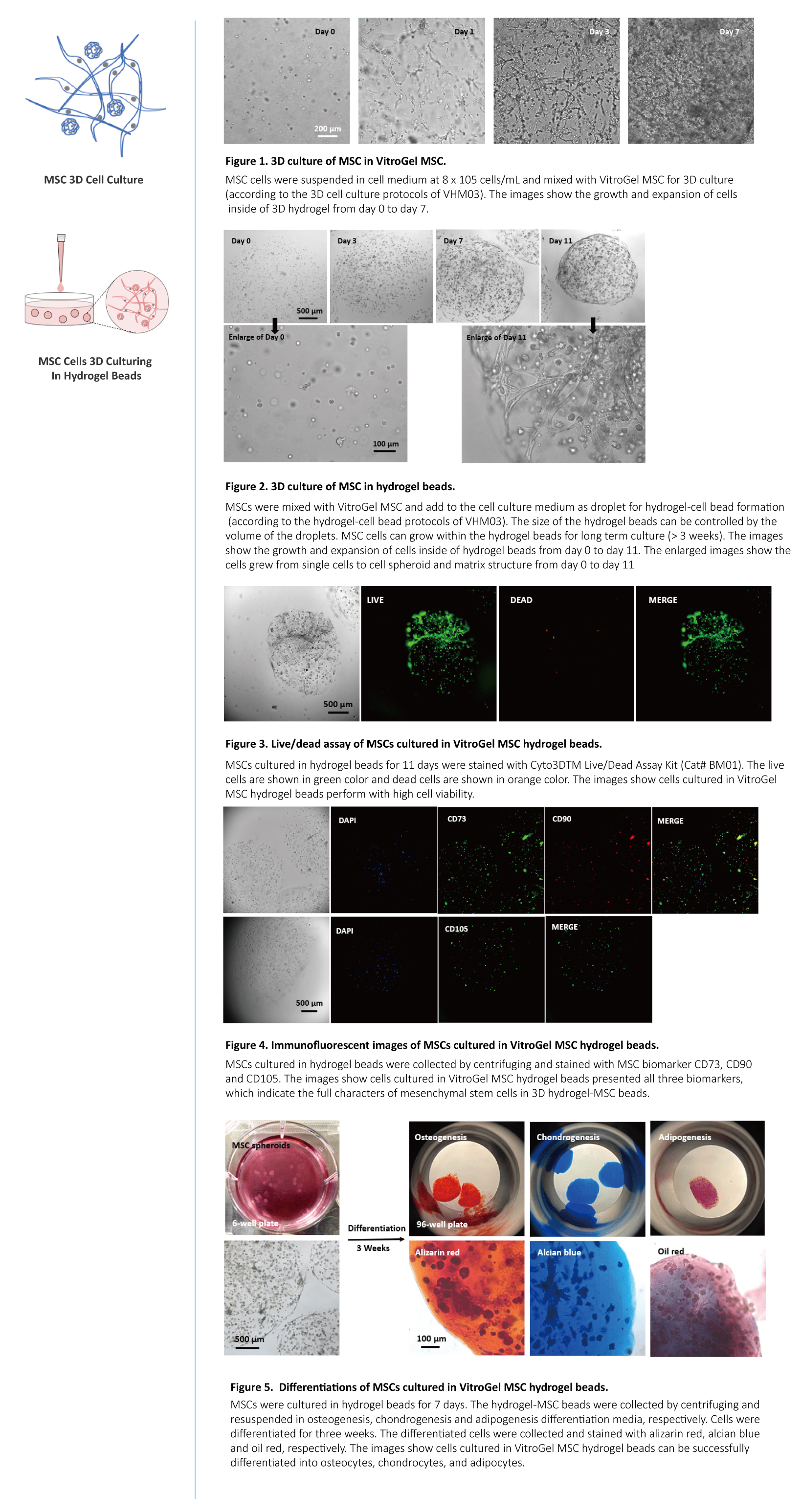
VitroGel® MSC is a xeno-free (animal origin-free) hydrogel system developed to support three-dimensional (3D) cultures of mesenchymal stem cells (MSCs). This hydrogel system can be used to make hydrogel cell beads for MSC scale-up. Microcarriers are not required for MSC scale-up.
VitroGel MSC is ready-to-use with an optimized formulation that fully supports the rapid expansion of MSCs. Cells directly thawed from liquid nitrogen or passaged from 2D culture vessels can be immediately mixed with the hydrogel solution for 3D culture or hydrogel-cell bead generation. This hydrogel system is compatible with most MSC culture media and tissue culture vessels. By using the VitroGel Cell Recovery Solution, the cell harvesting after 3D culture is simple and effective.
3D cell culture process in 20 min – “Just add cells”
VitroGel MSC is ready-to-use. Just mix with your cells.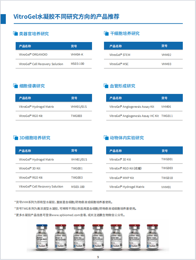
MSC 3D scale-up process in 20 min – Hydrogel-cell bead formation
VitroGel MSC is ready-to-use. Just mix with your cells and add as droplets.

Specifications
| Formulation | Xeno-free, functional hydrogel |
| Use | 3D cell culture, 2D hydrogel coating, hydrogel-cell bead formation |
| Operation | Ready-to-use at room temperature |
| Biocompatibility | Biocompatible, safe for animal studies |
| Injection | Injectable hydrogel for in vivo studies and lab automation |
| Cell Harvesting | Easy cell recovery using |
| pH | Neutral |
| Storage | Store at 2-8°C. Ships at ambient temperature |
| Sizes | 10 mL and 2 mL |
| Usage | 300 uses (10 mL), 60 uses (2mL) |
Data and References
ready-to-use, xeno-free (animal origin-free) hydrogel system for hPSCs 3D static suspension culture and scale-up
Overview
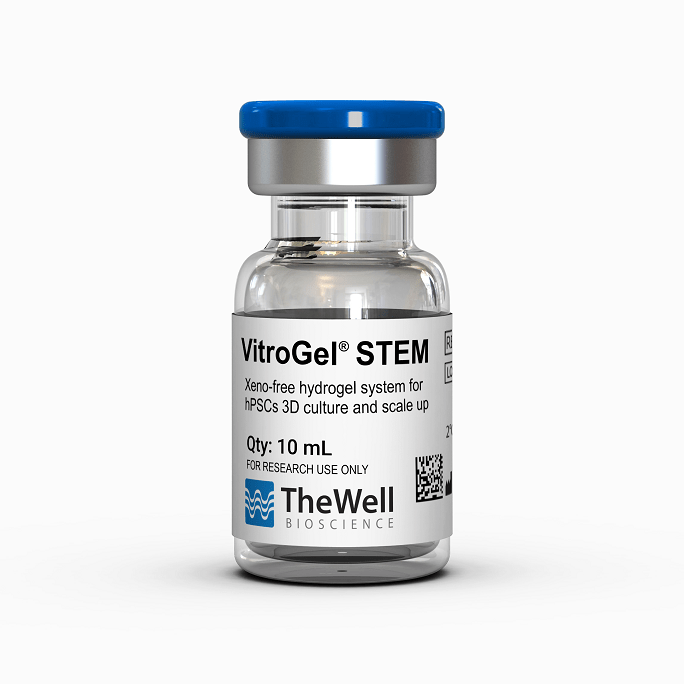
VitroGel® STEM is a xeno-free (animal origin-free) hydrogel system developed to improve the performance of three-dimensional (3D) static suspension cultures and scale-up of human pluripotent stem cells (hPSCs) to create a high-throughput system to model various tissue and disease states.
This hydrogel system is ready-to-use with an optimized formulation that fully supports the rapid expansion of high-quality 3D stem cell spheroids with pluripotent properties. hPSCs directly thawed from liquid nitrogen or passaged from 2D matrix coated culture vessels can be immediately mixed with the hydrogel solution for static suspension cultures. Moreover, the optimization protocol is ideal for time-sensitive experiments, as it does not require excessive medium exchanges, which can ultimately save on time and materials. This hydrogel system is compatible with most hPSC culture media and tissue culture vessels. Due to the unique static suspension culture procedure, the requirement for microcarriers for large-scale bioreactors is eliminated, making the cell harvesting simple and effective. The 3D stem cell spheroids that are developed using this system can be used for further sub-culturing, patterned differentiating, organoid developing, or re-establishing 2D culture morphologies.
Specifications
| Formulation | Xeno-free, functional hydrogel |
| Use | 3D static suspension culture for hPSCs |
| Operation | Ready-to-use at room temperature |
| Biocompatibility | Biocompatible, safe for animal studies |
| Injection | Injectable hydrogel for in vivo studies and lab automation |
| Cell Harvesting | 20 min cell recovery using |
| pH | Neutral |
| Storage | Store at 2-8°C. Ships at ambient temperature |
| Sizes | 10 mL and 2 mL |
| Usage | 10 mL hydrogel good for 90-180 mL suspension culture 2 mL hydrogel good for 15-30 mL suspension culture |
“Just add cells” 5 min protocol. No matrix coating required.
VitroGel STEM is ready-to-use. Just mix with your hPSCs. There is no laborous matrix coating required to maintain and expand your stem cells.

Advantages of the VitroGel STEM
Flexible
Undifferentiated stem cells can be easily transferred to 96 well plates, T-flasks, shaking flasks, and bioreactors, making them compatible with most culture vessels
Compatible with the majority of stem cell culture media
Easily switched back to 2D culture systems if needed
Easy to use
Easily seed stem cells directly from liquid N2 or 2D culture systems
Efficient cell harvesting or sub culturing without additional reagent
High performance
Can be used for stem cell scale-up with bioreactors at ultra-low agitating speed to maintain high cell viability and excellent cell growth rates
Yields high-quality 3D stem cells with full pluripotent properties
Cost-effective
Eliminates the need for matrix coating or microcarriers
Eliminates the need for shakers or stirrers for laboratory scale stem cell suspension cultures
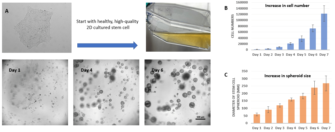 Figure 1. 3D static suspension culture of hPSC from 2D matrix culture
As shown in Figure 1, after 24 hours, small hPSC spheroids starts to form. From day 1 to 6, cells in the suspension cultures quickly grow, leading to the generation of healthy and high-quality stem cell spheroids. After day 3, cell number grow exponentially (Figure 1B) and spheroid size steadily increases (Figure 1C). The hPSC spheroids display characteristics of shallow craters or pockmarks, indicating expression of hPSC markers and successful expansion of healthy and high-quality stem cell spheroids. The resulting spheroids provide researchers with large numbers of healthy hPSCs for further experiments.
 Figure 2. 3D static suspension culture of hPSC directly from Liquid Nitrogen (LN2)
Start the suspension culture by using the healthy and high-quality cells directly from LN2. hPSC-hydrogel aggregates successfully to form healthy spheroids after 1 day in culture. The hPSC spheroids continue to expand from day 1 to 6 (Figure 2A). The resulting hPSC spheroids also show hallmark features of healthy and high-quality stem cell spheroids, i.e., shallow craters or pockmarks. Figure 2B shows that hPSC static suspension cultures from liquid nitrogen are positive for Alkaline Phosphatase, indicating successful expansion of healthy stem cell populations.
 Figure 3. Immunofluorescence images of hPSC spheroids with key pluripotent stem cell markers
VitroGel STEM ensures the undifferentiated state of stem cell lines during scale-up. As shown in Figure 3, hPSC aggregates in VitroGel STEM hydrogel and retain pluripotency after 7 days, evidenced by the expression of key pluripotent stem cell markers, SSEA4, OCT4, SOX2, and TRA-1-60.
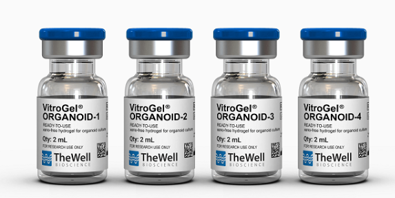
VitroGel® ORGANOID是专用于类器官培养的不含动物成分的水凝胶。支持来自患者来源样本的类器官、多能干细胞PSC来源、共培养、PDX模型的类器官生长。
VitroGel® ORGANOID
•专用于类器官培养的无异源成分水凝胶
•支持来自患者来源样品、干细胞、组织、共培养和 PDX 来源的各种类器官培养
•室温操作,自动化友好
•20 分钟轻松收获细胞(无酶)
VitroGel ORGANOID水凝胶在室温下可立即使用,具有pH值中性、透明、可渗透,与不同的成像系统兼容的特点。通过与细胞培养基简单混合,就可以形成水凝胶基质。适用于3D细胞培养和2D水凝胶包被。
可以通过将干细胞球从VitroGel STEM中转移到VitroGel ORGANOID水凝胶进行向类器官方向的分化,VitroGel ORGANOID水凝胶可以与VitroGel STEM(货号VHM02)联合使用,VitroGel STEM是一种用于3D静态悬浮培养和大规模扩增人iPS细胞的水凝胶系统。关键的生长因子和分子可以直接与水凝胶基质混合,或添加在水凝胶上面的培养基中。使用VitroGel细胞回收溶液可以轻松地回收在该系统中培养的类器官。
规 格
| 货号 | VHM04-K、VHM04-1、VHM04-2、VHM04-3 |
| 配方 | 无异源功能性水凝胶 |
| 应用 | 类器官培养 |
| 操作 | 在室温下即用型 |
| 生物相容性 | 生物相容性,对动物研究安全 |
| 注射 | 用于体内研究和实验室自动化的可注射水凝胶 |
| 细胞回收 | 类器官培养专用回收溶液 5-15 分钟回收细胞 |
| pH值 | 中性 |
| 储存 | 储存在2-8°C。 在环境温度下运输 |
| 规格 | 单个类器官水凝胶:10 mL Discovery Kit试剂盒:VitroGel ORGANOID-1、VitroGel ORGANOID-2、VitroGel ORGANOID-3、VitroGel ORGANOID-4 各2 mL |
使用 次数 | (10 mL):每孔 600 μL 使用 25 次或每孔 300 μL 使用 50 次 (2 mL):每孔120 μL 使用 25 次或每孔60 μL 使用 50 次 |
VitroGel ORGANOID Discovery Kit(货号VHM04-K)包括所有四种类型的类器官水凝胶(VitroGel ORGANOID-1、VitroGel ORGANOID-2、VitroGel ORGANOID-3、VitroGel ORGANOID-4),其配方中含有各种生物功基团、机械强度和可降解性,以满足不同类器官培养条件的需求。
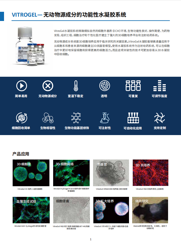
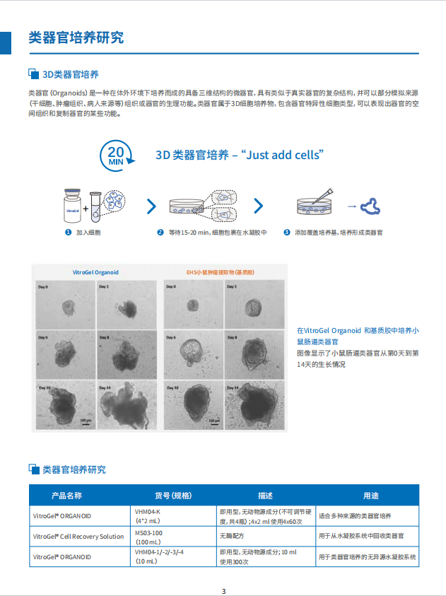
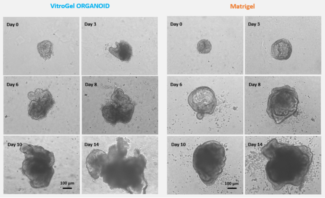
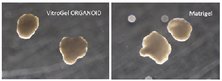
图1.在VitroGel Organoid和Matrigel基质胶中培养小鼠肠道类器官 从Day 0天到Day 14小鼠肠道类器官的生长情况
图2.用干细胞球培养人肠道类器官
液氮中取出iPSCs细胞接种于VitroGel STEM水凝胶进行静态悬浮培养(参考VHM02 protocol)。3-5天内形成了高质量干细胞球体具有完全的多能性(图中为SSEA4、OCT4、SOX2和TRA-1-60的阳性标记)。通过离心(100 g,3 min)收获干细胞球,并用VitroGel STEM重悬,在内胚层分化培养基中培养3天。然后通过离心(100g,3 min)收获内胚层细胞球,并用VitroGel STEM重悬,在中/后肠分化培养基中培养3-4天。通过离心(100 g,3 min)收集中/后肠胚,并用CDX2和E-Cadherin进行鉴定。用类器官培养基重悬中/后肠胚,并与VitroGel Organoid混合(参考VitroGel Organoid protocol)用于类器官形成和成熟后的长期培养。
产品操作指南
在VitroGel Organoid 和基质胶中培养小鼠肠道类器官
图像显示了小鼠肠道类器官从第0天到第14天的生长情况

在VitroGel Organoid 和Matrigel基质胶中培养脑类器官
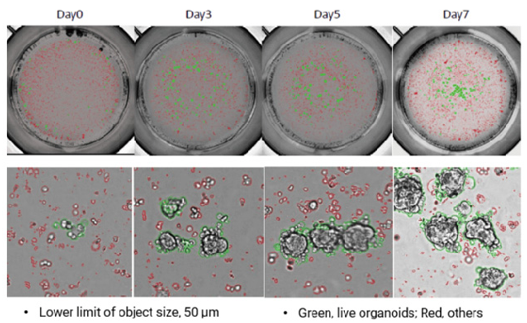
VitroGel Organoid 培养乳腺癌PDX类器官
VitroGel ORGANOID改善人胃类器官(HGO)和树突状细胞共培养模型中的免疫细胞-上皮相互作用。
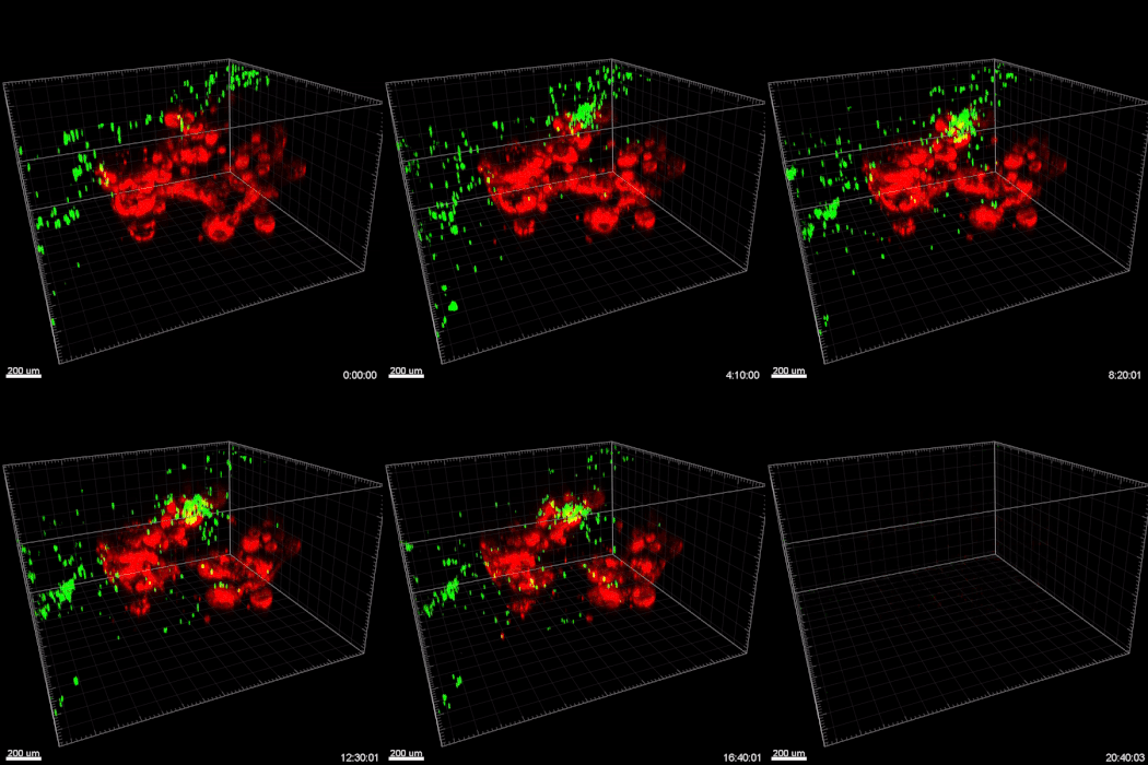
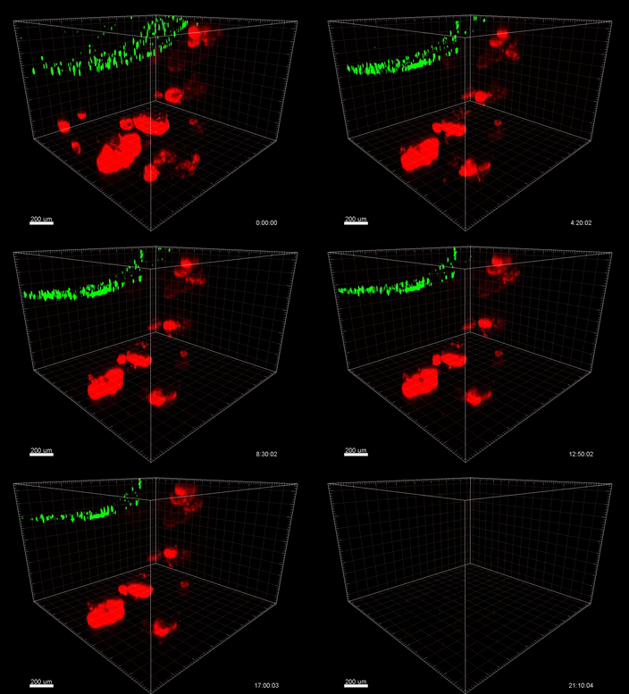
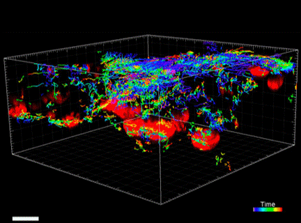
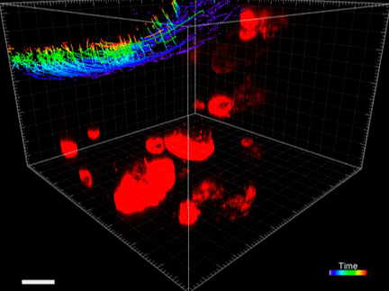
视频显示了在GOFlowChip上表达mCherry的人胃类器官,与CellTracker绿色标记的单核细胞来源的树突状细胞共同培养20小时。类器官嵌入VitoGel ORGANOID-3(左)和基质胶(右)中。与嵌入基质胶中的 HGO(红色)共培养时 MoDC(绿色)运动不良。改善MoDC(绿色)向嵌入VitroGel ORGANOID-3中的HGO(红色)的迁移。
(视频来源于蒙大拿州立大学的Barkan Sidar,Michelle Cherne,Jim Wilking和Diane Bimczok)。

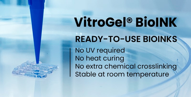
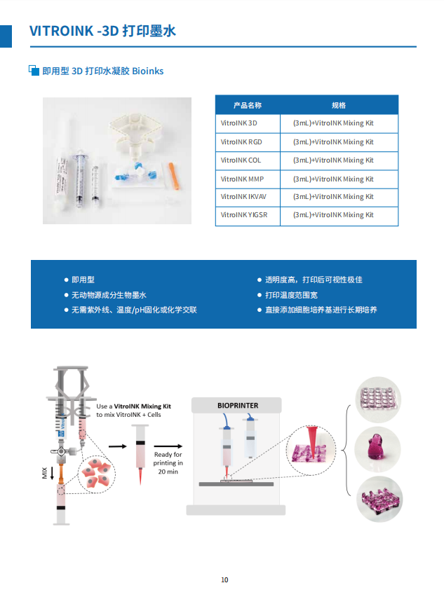
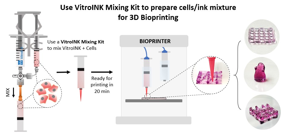
VitroINK™ are a new generation of bioink system that can print without UV or heat curing or chemical cross-linking.@ The VitroINKs are room temperature stable.@
Cells can be pre-mix in the hydrogel or directly mix with the bioink by using a dual syringe system.@ Multiple biological functional components can incorporate with this bio-ink for different applications.
Nozzle Tips

Application Note Nana-Fatima Haruna, Bhavya Shah,John Huang TheWell Bioscience, North Brunswick, NJ 08902 Introduction
There is an increasing need for the generation of data that accurately reflects the biological systems of patients 1-3. To achieve this goal,
researchers need to study disease models in microenvironments that depict the complex human extracellular matrix and support the
VitroGel™3D可用于3D细胞培养及2D包被,及其他灵活可变的应用.
3D细胞培养
VitroGel™3D可与细胞混合用于3D细胞培养,水凝胶基质支持细胞成长为3D克隆。可在水凝胶体系中加入药物或其他化合物,以进行药物筛选。
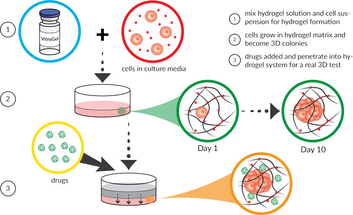
2D包被
VitroGel 3D可用于包被培养皿表面以促进细胞生长,不同的硬度及质地会导致不同的细胞行为。同时,随着时间的推移,细胞可能进入水凝胶内部,可做细胞侵袭研究。
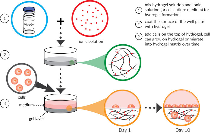
其他研究
以适当比例(推荐4:1 v/v)混合水凝胶和细胞悬液(或其他离子溶液),可以形成可注射的水凝胶,用于异种移植,药物递送,生物打印及微列阵等。
另外,可根据不同的物理/化学特性和成胶次数调节水凝胶的特性及制备方案,也可通过调节水凝胶溶液的浓度来控制水凝胶强度。可以通过这些灵活可调节性,进行多种应用。
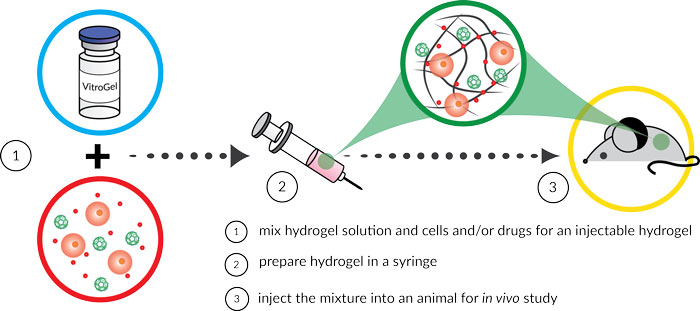
VitroGel 3D为细胞创造一个功能性和优化的环境,让细胞感觉在家一样。从2D包被,3D细胞培养到动物注射,VitroGel 3D让利用相同系统进行体内体外研究成为可能。
用户使用VitroGel 3D系统主要用于研究癌症和肿瘤细胞生物学,干细胞和组织工程,药物发现和毒性研究等方面。
下表列出了已在VitroGel 3D系统中成功测试的细胞系。可根据不同的细胞系和培养基组分优化VitroGel 3D水凝胶系统,以得到更好的结果。推荐培养不同的细胞系时,都进行水凝胶浓度梯度测试。
Tables of successful cell types with Vitrogel 3D (TWG001)
| CELL TYPES | APPLICATIONS | CULTURE MEDIUM | DILUTION |
|---|---|---|---|
| 4T1 cells | 3D culture | RPMI 1640 with 10% FBS | 1:2 |
| BL5 human beta cells | 2D and 3D culture | DMEM with 10%FBS | 1:3 |
| CD8+ T cells | 3D culture | RMPI 1640 with 10% FBS | 1:3 |
| HeLa cells | 3D culture | DMEM with 10%FBS | 1:3 |
| Human Nthy-ori 3-1 cells | 3D culture | RPMI 1640 with 10% FBS | 1:3 |
| KHOS cells | 3D culture | RPMI 1640 with 10% FBS | 1:1 to 1:3 |
| MDA-MB-231 cells | 3D culture | RPMI 1640 with 10% FBS | 1:2 |
| Priess human lymphoblastoid cells | 2D and 3D culture | RPMI 1640 with 10% FBS | 1:3 |
| Red Blood Cells | 3D culture | Alsever's solution | 1:1 to 1:3 |
| T47D cells | 3D culture | RPMI 1640 with 10% FBS | 1:2 |
| U2OS cells | 3D culture | RPMI 1640 with 10% FBS | 1:1 to 1:3 |
Tables of successful cell types with Vitrogel 3D-RGD (TWG002)
| CELL TYPES | APPLICATIONS | CULTURE MEDIUM | DILUTION |
|---|---|---|---|
| 4T1 cells | 3D culture | RPMI 1640 with 10% FBS | 1:2 |
| A549 cells | 3D culture | DMEM with 10% FBS | 1:1 to 1:3 |
| Au565 cells | 3D culture | RPMI 1640 with 10% FBS | 1:3 |
| Beta TC3 cells | 3D culture | DMEM with 10% FBS | 1:3 |
| BT 474 cells | 3D culture | DMEM with 10% FBS | 1:3 |
| DLD1 cells | 3D culture | DMEM with 10% FBS | 1:1 to 1:3 |
| Fuji cells | 3D culture | RPMI 1640 with 10% FBS | 1:1 to 1:3 |
| H460 cells | 3D culture | RPMI 1640 with 10% FBS | 1:1 to 1:3 |
| HCT 116 cells | 2D and 3D culture | McCoys 5 with 10% FBS | 1:1 |
| HCT-8 cells | 3D culture | RPMI 1640 with 10% FBS | 1:3 |
| HEK 293 cells | 3D culture | DMEM with 10% FBS | 1:3 |
| Hela cells | 2D and 3D culture | DMEM with 10% FBS | 1:3 |
| HepG2 cells | 3D culture | DMEM with 10% FBS | 1:1 to 1:3 |
| Human iPSCs | 2D and 3D culture | mTeSR1 | 1:3 |
| Ins-1 Cells | 3D culture | RPMI 1640 with 10% FBS | 1:3 |
| MCF-12A cells | 3D culture | DMEM/F-12 with 10% FBS | 1:3 |
| MDA-MB-231 cells | 3D culture | L-15 medium with 10% FBS | 1:1 to 1:3 |
| Melanoma cells | 3D culture | RPMI 1640 with 10% FBS | 1:1 to 1:3 |
| OVCAR-3 cells | 3D culture | RPMI 1640 with 10% FBS | 1:1 to 1:3 |
| Panc-1 cells | 3D culture | DMEM with 10% FBS | 1:1 to 1:3 |
| SKB3 cells | 2D and 3D culture | DMEM with 10% FBS | 1:1 to 1:3 |
| SYO-1 cells | 3D culture | RPMI 1640 with 10% FBS | 1:1 to 1:3 |
| T47D cells | 3D culture | RPMI 1640 with 10% FBS | 1:3 |
| UD 145 cells | 3D culture | EMEM with 10% FBS | 1:1 to 1:3 |
Tables of successful cell types with Vitrogel 3D RGD-PLUS (TWG003)
| CELL TYPES | APPLICATIONS | CULTURE MEDIUM | DILUTION |
|---|---|---|---|
| MCF-7 cells | 2D and 3D culture | DMEM with 10% FBS | 1:1 to 1:3 |
| MCF-12A cells | 3D culture | DMEM/F-12 with 10% FBS | 1:3 |
| MDA-MB-231 cells | 3D culture | L-15 medium with 10% FBS | 1:1 to 1:3 |
| OP9 cells | 2D and 3D culture | Alpha-MEM with 10% FBS | 1:3 |
| Panc-1 cells | 2D and 3D culture | DMEM with 10% FBS | 1:1 to 1:3 |
| SF 268 cells | 2D and 3D culture | RPMI 1640 with 10% FBS | 1:1 to 1:3 |
| SF 295 cells | 2D and 3D culture | RPMI 1640 with 10% FBS | 1:1 to 1:3 |
| SNB 75 cells | 2D and 3D culture | RPMI 1640 with 10% FBS | 1:1 to 1:3 |
| U-87 MG cells | 2D and 3D culture | EMEM with 10% FBS | 1:1 to 1:3 |
| U-251 MG cells | 2D and 3D culture | EMEM with 10% FBS | 1:1 to 1:3 |


