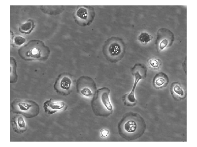
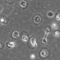
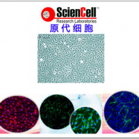
The cells were first observed by Karl Wilhelm von Kupffer in 1876. The scientist called them "Sternzellen" (star cells or hepatic stellate cell) but thought, inaccurately, that they were an integral part of the endothelium of the liver blood vessels and that they originated from it. In 1898, after several years of research, Tadeusz Browicz identified them, correctly, as macrophages.
Kupffer cells, also known as stellate macrophages and Kupffer-Browicz cells, are specialized macrophages located in the liver lining the walls of the sinusoids that form part of the reticuloendothelial system (RES) (or mononuclear phagocyte system).
| Product | Catalog no. | Amount | Storage |
| Hepatic Kupffer Cell | HKC001 | 1X10^6/vial | in liquid nitrogen |
Product Use
For Research Use Only.Not for use in diagnostic procedures
Culture Conditions
Culture Type:Adherent
Temperature Range:36℃ to 38℃
Incubator Atmosphere:Humidified atmosphere of 5% CO2
Size:The typical hepatocyte is cubical with sides of 20-30 µm
Passaging Adherent Cells
All solutions and equipment that come in contact with the cells must be sterile.
Always use proper sterile technique and work in a laminar flow hood.
1. Remove and discard the spent cell culture media from the culture vessel.
2. Wash cells using a balanced salt solution without calcium and magnesium (approximately 2 mL per 10 cm2 culture surface area). Gently add washsolution to the side of the vessel opposite the attached cell layer to avoiddisturbing the cell layer,and rock the vessel back and forth several times.
Note: The wash step removes any traces of serum, calcium, and magnesium that would inhibit the action of the dissociation reagent.
3. Remove and discard the wash solution from the culture vessel
4. Add the pre-warmed dissociation reagent such as trypsin or TrypLE™to the side of the flask; use enough reagent to cover the cell layer (approximately0.5 mL per 10 cm2). Gently rock the container to get complete coverage of the cell layer.
5. Incubate the culture vessel at room temperature for approximately 2minutes.
Note: that the actual incubation time varies with the cell line used.
6. Observe the cells under the microscope for detachment. If cells areless than 90% detached, increase the incubation time a few more minutes, checking for dissociation every 30 seconds. You may also tap the vessel to expedite cell Tetachment.
7. When ≥ 90% of the cells have detached, tilt the vessel for a minimallength of time to allow the cells to drain. Add the equivalent of 2 volumes (twice thevolume used for the dissociation reagent) of pre-warmed complete growth medium.Disperse the medium by pipetting over the cell layer surface several times.
8. Transfer the cells to a 15-mL conical tube and centrifuge then at200 × g for 5 to 10 minutes. Note that the centrifuge speed and time vary based on the cell type.
9. Resuspend the cell pellet in a minimal volume of pre-warmed complete growth medium and remove a sample for counting.
10. Determine the total number of cells and percent viability using a hemacytometer, cell counter and Trypan Blue exclusion, or the Countess® Automated CellCounter. If necessary, add growth media to the cells to achieve the desired cellconcentration and recount the cells.

