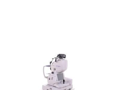
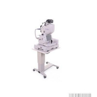
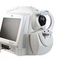
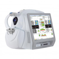
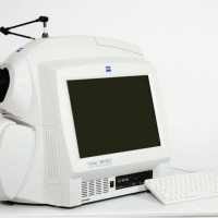
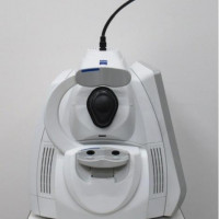
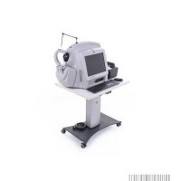
Reconditioned (used),
Technical condition: very good,
Visual condition: very good,
Real photos of the product,
Made in Japan, (2012),
Power supply: 230 V,
Frequency: 50/60 Hz,
Power: 200 VA (max. 400 VA),
Software: 3D OCT - version 8.43,
TOPCON 3D-OCT 2000 combines a fundus camera generating high-quality color images with a state-of-the-art optical spectral tomography OCT,
The high scanning speed (50,000 A-scans/s), and high resolution (5µm) allow building spatial images of the examined tissues on which even the smallest changes can be observed,
Thanks to noise-reducing algorithms and Infrared/3D tracking technology, OCT sections perfectly illustrate the structure of the vitreous body and retina,
The device provides a variety of tools that allow segmentation and extraction of layers, cropping, smoothing, and moving them,
The PinPoint Registration function allows the operator to mark the finest details in the area of the taken fundus image and observe them in the OCT presentation on 2D and 3D scans or thickness maps,
A built-in fundus camera with a high-quality digital camera (16.2 Mpx) complements OCT diagnostics, allowing to visualize the eye structure in detail and revealing any pathological changes,
Various analytical functions combined with color photography and 3D OCT imaging enable imaging and analysis of the cornea and anterior segment of the eye,
Tool
Diagnostic tools are complemented by such functions as distance measurement, curvature measurement, and seepage angle measurement,
TOPCON 3D OCT-2000 technical specifications:
Light source: SLD,
Wavelength: 840 nm,
Retinal scanning field /multiplicity: 45°,
Axial resolution: 5; 6 µm,
Transverse resolution: 20 µm,
Max. scanning depth: 2.3 mm,
Integration with funduscamera,
Ability to analyze structures of the anterior segment of the eye,
Visualization of a cross-section through a selected section of the fundus of the eye,
3D visualization,
Wide scan of 12 x 9 mm,
Built-in fixation point,
The tomograph is equipped with a NIKON D90 camera,
Matrix: CMOS, APS-C (23.6 x 15.8 mm),
Matrix resolution: 12 Mpix,
Maximum image size: 4288 x 2848,
Recording format: Motion JPEG,
Video resolution:
1280 x 720 (24 fps),
640 x 424 (24 fps),
320 x 216 (24 fps),
Tomograph placed on a mobile 4-wheel TOPCON ATE-600 table (2 braking wheels) with an adjustable range of 68 - 82 cm,
Tomograph equipped with additional instrumentation placed on an additional 4-wheel mobile table (2 braking wheels):
AIO computer:
Built-in screen: 21
